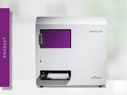
PHERAstar FSX
Powerful and most sensitive HTS plate reader


Learn about AlphaScreen technology—an innovative, no-wash assay platform for biomolecular interactions, screening, and drug discovery.
AlphaScreen® (ALPHA for Amplified Luminescent Proximity Homogeneous Assay) is bead-based chemistry used to study interactions between molecules in a microplate. Originally developed as a methodology for diagnostic assays called LOCI® (Luminescent Oxygen Channeling Immunoassay) in 19941, today the AlphaScreen Technology (Alpha Technology) includes AlphaScreen, AlphaLISA®, and AlphaPlexTM. It is mainly used in the life sciences for screening purposes.
The fundamental principle of the AlphaScreen technology relies on binding two different molecules of interest to specific beads. In case of interaction between the two molecules and the resulting proximity of the two beads, an energy transfer from one bead to the other takes place. This results in the production of a chemiluminescent signal. The AlphaScreen technology is mainly used in high-throughput screening assays to assess biomolecular interactions, the formation/depletion of a substrate or product, post-translational modifications and to quantify analytes.
Besides interaction assays (including ligand/receptor, protein/protein, protein/DNA), the AlphaScreen technology can also be applied to GPCR functional assays (second messenger detection), enzymatic assays, and immunoassays.
AlphaScreen assays are based on two types of hydrogel-coated beads, called donor and acceptor beads. The two bead types contain different chemicals that are key to the generation of the luminous signal. The donor bead contains a photosensitizer, which upon excitation by light at 680 nm, converts oxygen (O2) into an exciting form, singlet oxygen (1O2). Singlet oxygen molecules have a reduced lifetime (4 microseconds half-life) and can diffuse approximately 200 nm in solution before falling back to the ground state.
In the absence of acceptor beads, the singlet oxygen molecules fall back to the ground state without producing any light signal. In case an acceptor bead is within 200 nm, energy is transferred from the singlet oxygens to the bead, resulting in light production. AlphaScreen acceptor beads contain three chemical dyes, thioxene, anthracene, and rubrene. Singlet oxygen molecules initially react with thioxene to generate light. This is transferred to anthracene and to rubrene and results in broad light emission from 520 nm to 620 nm.2 The half-life of the signal decay reaction is 0.3 seconds.
In binding assays, one binding partner (e.g. a receptor) is linked to the donor, while the other (e.g. a ligand) to the acceptor beads. When receptor and ligand interact, chemical energy is transferred from one bead type to the other and a luminous signal is produced (fig. 1). AlphaScreen can also be employed for competition or cleavage assays. In these cases, a reduction in signal intensity is measured.
Donor beads are typically available as streptavidin conjugates, exploiting the specific binding of biotin to streptavidin to label biomolecules. Instead, acceptor beads are primarily conjugated to antibodies. Hence, the second binding partner usually requires the presence of a corresponding antigen in order to be bound (fig. 2). Alternatively, both bead types can be directly coated with the binding partners by reductive amination.
As both types of beads are around 250 nm in diameter, they are too small to sediment in biological buffers. In addition, they do not clog tips or injectors and can be easily managed by automated liquid handling equipment. Yet, they are large enough to be filtered or centrifuged. The hydrogel coating allows conjugation of biomolecules to the beads while reducing non-specific binding and self-aggregation.
AlphaLISA
AlphaLISA is a further development of the AlphaScreen technology that relies on the same donor beads but uses a different type of acceptor beads. In AlphaLISA beads, anthracene and rubrene are substituted by europium chelates. Excited europium emits light at 615 nm with a much narrower wavelength bandwidth than AlphaScreen (fig. 3). Hence, AlphaLISA emission is less susceptible to compound interference and can be employed for the detection of analytes in biological samples like cell culture supernatants, cell lysates, serum, and plasma.
AlphaLISA allows the quantification of secreted, intracellular, or cell membrane proteins. For biomarker detection, AlphaLISA is mainly employed as a sandwich immunoassay. A biotinylated anti-analyte antibody binds the streptavidin donor bead while a second anti-analyte antibody is conjugated to AlphaLISA acceptor beads. In the presence of the analyte, beads come into close proximity. Donor bead excitation releases singlet oxygen molecules that transfer energy to the acceptor beads with light emission at 615 nm (fig. 4). Alternatively, competition immunoassays can also be adapted.
Compared to classical ELISAs, AlphaLISA is said to provide better sensitivity, a wider dynamic range, and robust performance with reduced assay times. Moreover, since it does not require washing steps (see below), it is easily adaptable to automated high-throughput screening.

AlphaPLEX
AlphaPlex is a further development that allows quantification of up to three analytes in a single well by using different acceptor beads that emit at distinct wavelengths. In these beads, anthracene and rubrene are substituted by either terbium chelates (for AlphaPlex 545) or samarium chelates (for AlphaPlex 645). Biotinylated anti-analyte antibodies bind the streptavidin donor beads. Corresponding anti-analyte antibodies are conjugated to either AlphaPlex or AlphaLISA acceptor beads. In the presence of the target analytes, excitation of the donor beads and the consequent release of singlet oxygen triggers chemiluminescent light emission from the different acceptor beads.
Each type of bead emits light at a different wavelength. In addition to 615 nm for AlphaLISA beads, the other emission peaks are at 545 nm for terbium beads and at 645 nm for samarium beads (fig. 5). Accordingly, the development of triplex assays is enabled by the use of AlphaLISA, AlphaPlex 545, and AlphaPlex 645 acceptor beads in one assay.2
The measurement of the AlphaScreen chemistry is predominantly performed on microplate readers. As it is an independent read mode, assay detection requires a microplate reader with AlphaScreen detection mode. This includes detection capabilities for AlphaLISA and AlphaPlex as well. As this technology is often used in high-throughput drug screening, plate readers should be compatible with 384 and 1536 well plates.
The AlphaScreen plate reader basic setup consists of a light source, excitation and emission filters for wavelength selection, and a photomultiplier tube (PMT) detector.
Light source: xenon lamp or laser
As the photosensitizer on donor beads is specifically excited at 680 nm, a xenon flash lamp or a specific excitation laser are used as light source. A dedicated AlphaScreen laser focuses more energy at 680 nm, outperforming xenon lamps and leading to better results with a broader dynamic range and increased signal-to-noise ratios.
Dedicated AlphaScreen excitation lasers are commonly available on high-end as well as HTS-dedicated multi-mode microplate readers like the PHERAstar FSX and CLARIOstar Plus. Budget readers are usually equipped with a xenon flash lamp for AlphaScreen detection.
AlphaPLEX entails the detection of different emission wavelengths. Although these can be measured sequentially, simultaneous detection of two emissions ensures both data quality and speed advantages, especially for screening assays. Accordingly, Simultaneous Dual Emission detection helps increase throughput with dual emission AlphaLISA-AlphaPlex assays.
Cross-talk reduction
Cross-talk is the light from any but the measured well that is unspecifically measured by the detector and negatively affects the signal of the measured well.
Since the light produced in a well by an AlphaScreen reaction is diffuse, it can spread to neighboring wells and directly to the site of detection, even though another well is measured. This leads to biased signals, higher signal variations, and lower overall sensitivity.
Undesired light signals reach the detector either from above the plate and/or through the wall of the wells and need to be differently addressed (fig. 6).
Both cross-talk reduction tools are available on the BMG LABTECH plate readers PHERAstar FSX and CLARIOstar Plus.
AlphaScreen/AlphaLISA detection can be performed on the PHERAstar FSX and CLARIOstar Plus. Since a high-intensity excitation source is beneficial to the assay, the CLARIOstar and PHERAstar FSX have dedicated, solid-state lasers which produce high-intensity light at 680 nm. In addition, the PHERAstar FSX offers specific AlphaPlex optic modules that allow the simultaneous measurement of two Alpha signals.
AlphaScreen provides a homogeneous, sensitive, and down scalable platform that is specifically well-suited for high-throughput screening applications.
High signal-to-background ratio
The signal generation process is a cascade reaction that produces a high signal amplification. This results in high sensitivity. In addition, the background is low. This is mainly the consequence of the excitation wavelength being in the red range and at a higher wavelength than the emission signal. This reduces the auto-fluorescence generated by biomolecular components that is usually excited in the blue-green range. Moreover, the time delay between excitation and emission further eliminates auto-fluorescent noise.
Homogeneous assay
The AlphaScreen chemistry is homogeneous: detection of the bound donor-acceptor bead complex does not require physical separation from the unbound components to reduce the background. Consequently, it does not necessitate in-between separation or washing steps and can be executed with a simple add-and-read protocol. This minimizes handling steps and makes it more convenient and less time-consuming than other methods. Hence, it is particularly suited for automation-supported screening purposes. This is one of the reasons for its large adoption in drug discovery and HTS.
Downscaling and miniaturization
Due to its high sensitivity and low background, AlphaScreen is easily adaptable to 1536-well plates and can be easily downscaled to a few µl with no change in reagent concentration or assay robustness as shown in the application note “Miniaturization of a cell-based TNF-α AlphaLISA assay”.
Appropriate microplate and handling
White microplates are best suited for AlphaScreen measurements as they reflect the light signal. Black plates absorb light and lead to both reduced signal and background. Grey plates offer the best compromise between cross-talk reduction and signal reflection. Detailed information on the plate choice is found in our blog post: "The microplate: utility in practice."
As white plates have an intrinsic phosphorescence, they emit light if exposed to sun or room light. This unspecific signal is measured by the reader together with the light emitted by the sample and results in increased blanks and background, and a reduced assay window. It is therefore recommended to either prepare white plates in the dark or to leave them in the dark for approximately 15 min before measuring.
Environment lighting
AlphaScreen chemistry is light sensitive. Ideally, it should be prepared and run under subdued light conditions (under 100 Lux). As this is not always possible, an enclosed room should be outfitted with green filtered lighting. Light fixtures in the immediate proximity of the workspace and/or plate reader should be covered with green filters. The use of green filters has been shown to be almost as effective as running the assay in the dark (fig. 7).
Plates to be incubated for extended periods should be protected from light - covered with a dark cloth, aluminium foil, or a cardboard box. Avoid exposing the microplate or plate reader to direct sun or intense light.
Environment temperature
The AlphaScreen chemistry is sensitive to temperature. As such, dramatic room temperature fluctuations should be avoided as they negatively affect signal generation, intensity, and stability. Typically, AlphaScreen signal variation is around 8% per °C.3
To avoid signal intensity gradients across the plate while being read, reagents and microplates should also be equilibrated with the room temperature. For plates, this can be easily achieved by incubating them in a THERMOstar incubator. The PHERAstar FSX with AAS system even allows to actively set the reader to any temperature between 18° and 45°C and can thereby cool the incubation chamber of the device to room temperature or below. In this application note, the AAS system’s ability to maintain a stable temperature, reduce evaporation and the cool-down time from high to low temperatures is demonstrated.
Cell culture media and sera
Cell culture media like RPMI 1640, MEM, and DMEM negatively affect signal intensity. The presence of a 10% culture medium results in about 30% signal quenching. Similarly, 10% foetal calf serum reduces the signal by about 25%.3 Rinsing cell samples with an appropriate buffer like PBS before applying the AlphaScreen chemistry is therefore recommended.
The AlphaScreen technology can be applied to analyse binding or biochemical events in 96, 384, and 1536 well plate formats for different biological settings. The technology is particularly diffused in the drug screening community.
AlphaScreen is applied in different assays like GPCR functional assays (cAMP, IP3, and phospho-ERK1/2), enzymatic assays (tyrosine kinase, helicase, protease, phosphatase), interaction assays (cytokine binding assays, nuclear receptor functional assays, ligand-receptor binding assays, protein/protein, protein/DNA, protein/peptide), ELISA-like immunoassays for analyte quantification (TNF-α, IgG).
Binding assay: protein-peptide interaction and inhibitor identification
In the application note PHERAstar measures AlphaScreen assay to develop selective inhibitors for the human YEATS domains, we show an example of an AlphaScreen-based interaction assay.
YEATS domains are epigenetic regulators. The YEATS-containing ENL has been confirmed as a major driver of several types of acute leukaemia. ENL is therefore a drug discovery target. To screen for selective inhibitors, a histone 3 (H3) – YEATS domain binding assay was set up using the AlphaScreen Histidine detection kit. The donor bead is coupled via streptavidin to acylated H3 and the acceptor bead to the YEATS domain. When protein and peptide interact, the different beads are brought together and an AlphaScreen signal is generated (fig. 8). Inhibitors reduce the emission signal by disrupting the H3-YEATS interaction.
Immunoassay: phospho-ERK1/2 detection
Besides interaction, immunoassays can also be implemented. Responses for many GPCRs are not easily measured in current assay formats. The ERK phosphorylation cascade results in the phosphorylation of both cytoplasmic and nuclear proteins regulating gene transcription. Hence it can be used to screen for cellular changes induced by GPCR engagement.
For this purpose, the AlphaScreen SureFire® ERK1/2 phosphorylation assay was used on cell lysates. This is a sandwich immunoassay. A phosphorylated cellular protein is sandwiched between an anti-analyte antibody associated with a streptavidin-coated donor bead and an anti-phospho antibody associated with a Protein A-conjugated acceptor bead (fig. 9). Phosphorylation of the analyte results in an increase in the light signal. This is shown in the application note An AlphaScreen SureFire Phospho-ERK1/2 assay.
AlphaScreen, AlphaLISA, AlphaPlex, and SureFire® are registered trademarks of PerkinElmer.
Powerful and most sensitive HTS plate reader
Most flexible Plate Reader for Assay Development