Introduction
Phagosome maturation allows internalized particles, such as bacteria and apoptotic cells, to be trafficked to acidified compartments, leading to proteolytic degradation of unwanted cargo1. Investigating molecular dynamics of phagosome maturation is essential to fully understand mechanisms of the innate immune response to infection and in diseases of particulate matter such as gout2. In recent years, there has been an increased interest in realtime fluorescent cell-based assays to study the kinetics of phagosome maturation. This allows the investigation of phagosomal pH changes as well as proteolysis of phagocytosed particles3,4. Plate readers offer a much more rapid and automated read-out of fluorescence compared to flow cytometry and confocal microscopy, allowing for dynamics to be followed closely over time.
Assay principle
Carboxylated silica microspheres can be conjugated to succinimidyl ester fluorescent reporters of phagosomal pH e.g. pHrodo Red. Following uptake by macrophages, pHrodo Red emits fluorescence when encountering an acidic environment (Fig 1). pHrodo Red signal is read over time to follow phagosome maturation in real time.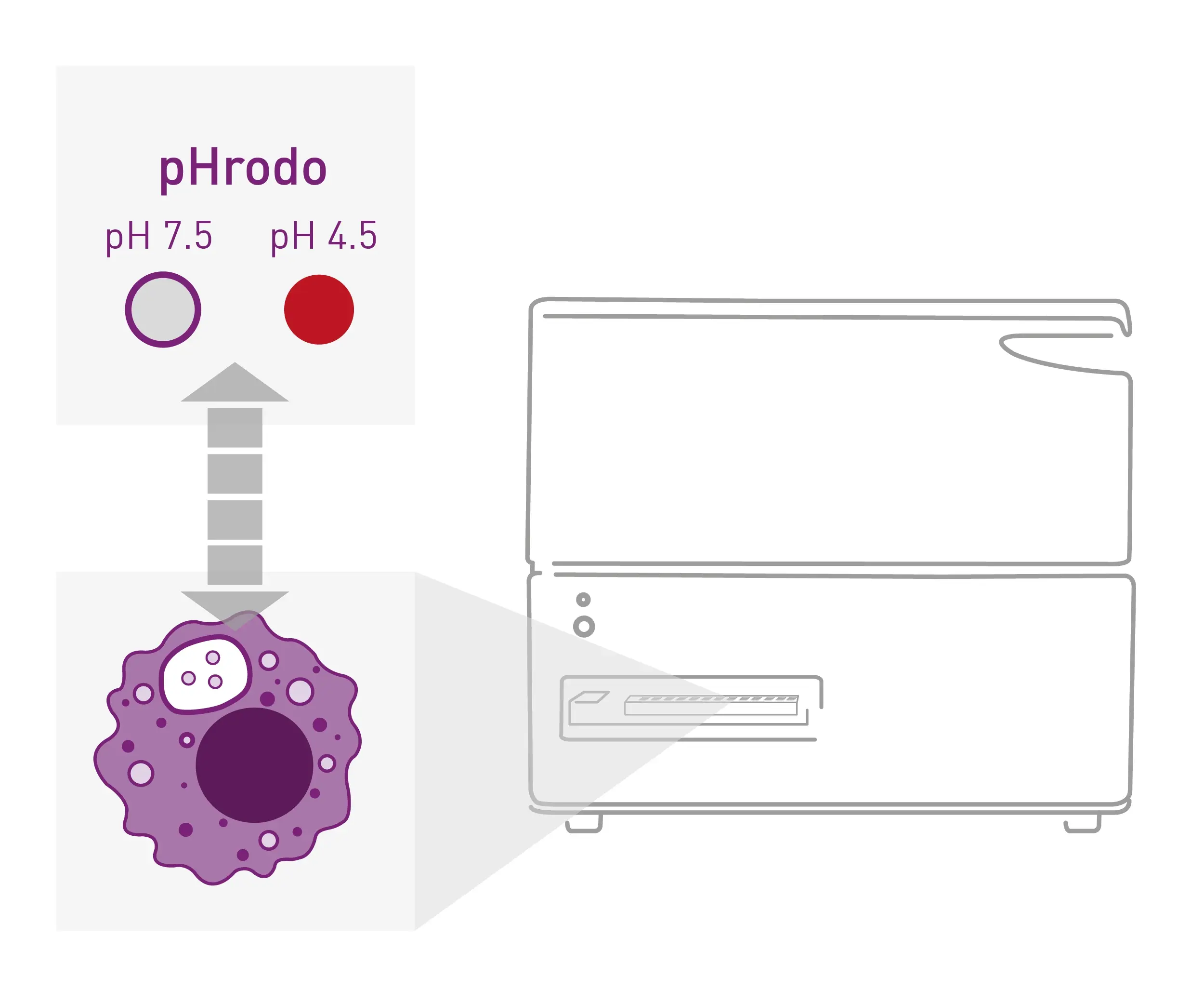
- Carboxylated silica beads (#PSI-3.0, Kisker Biotech), pHrodo Red, SE (#P36600, Thermo Fisher Scientific)
- Alexa Fluor 488 NHS Ester (#A20100, Thermo Fisher Scientific)
- Assay buffer: Phenol Red free DMEM (#21063029, Gibco), 5% FBS
- 96 well black/clear bottom plate, TC treated (#732-2604, Thermo Fisher Scientific)
- BMG CLARIOstar Plus (BMG LABTECH)
Experimental procedure
Carboxylated silica beads were coupled to pHrodo Red and Alexa Fluor 488 esters (to standardise for uptake) following manufacturer’s instructions [3].
1x10⁵ RAW 264.7 cells were plated per well in DMEM 10% FBS 1% P/S and allowed to adhere overnight. Cells were pre-treated with relevant inhibitors or vehicle control for 30 min prior to bead uptake. Media was replaced with assay buffer (no bead control) or bead slurry (diluted 1:300 in assay buffer) and incubated for 5 min at RT. Beads were washed out with assay buffer and inhibitors were replaced.
Instrument settings
|
Optic settings
|
Fluorescence, plate mode kinetic
|
|
|
Monochromator, bottom optic
|
||
|
AF488
|
Ex: 488-14
Em: 535-30 |
|
|
pHrodo
|
Red Ex: 550-20
Em: 605-40 |
|
|
Focal height
|
2.5
|
|
|
General settings
|
Number of flashes | 51 |
|
Scan settings
|
Spiral averaging | |
| Scan diameter | 4 mm | |
| Kinetic settings | Number of cycles | 120 |
| Cycle time | 60 s | |
| Shaking | Before first cycle, double orbital,100 rpm, 30 s | |
|
Incubation
|
37ºC, 5% CO2 |
|
Results & Discussion
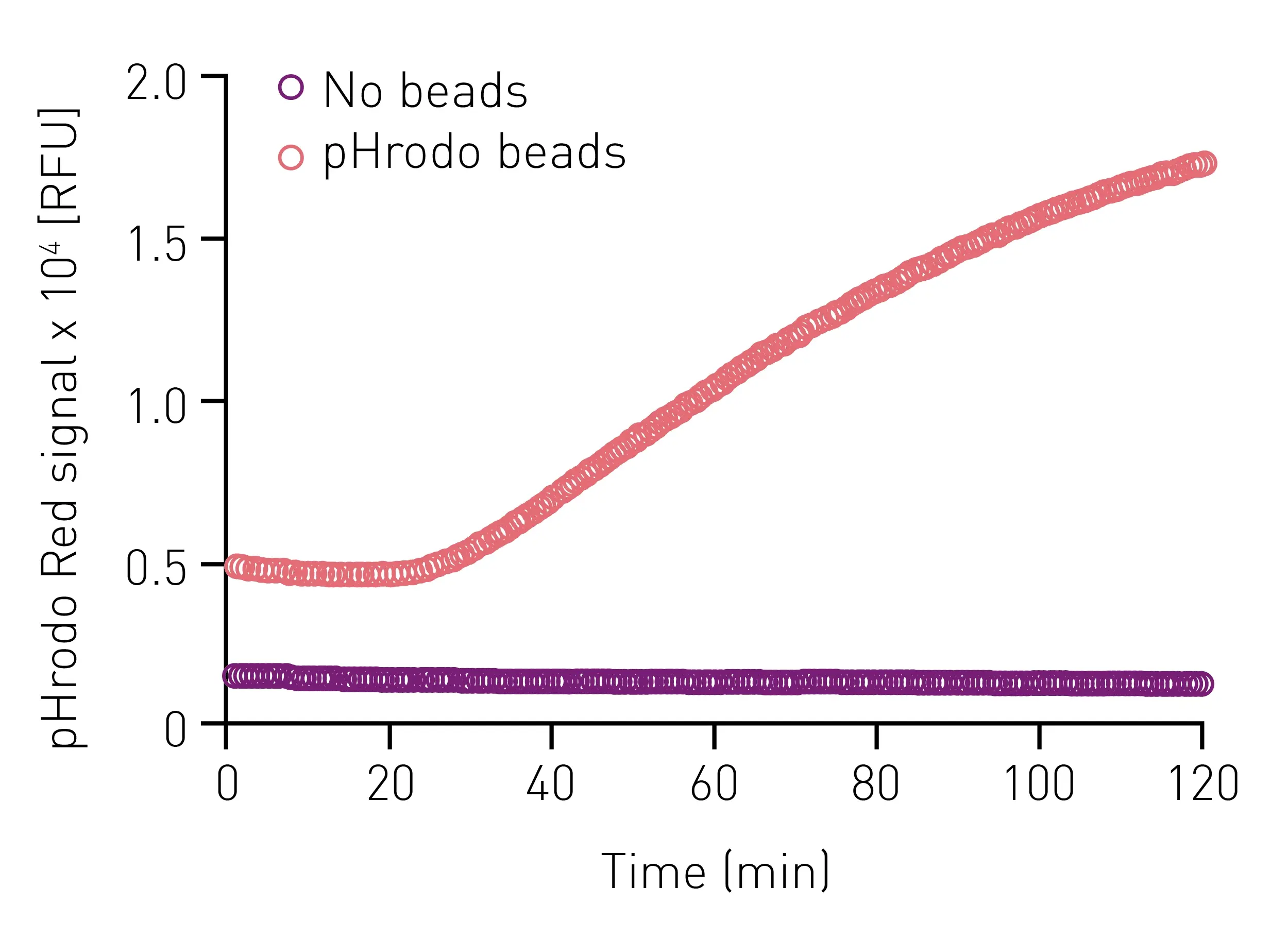
Using the described settings, increasing pHrodo Red fluorescence over time, following bead uptake, was able to be detected (Fig 2). As a control, cells that were treated with assay buffer in the absence of beads showed no increase in fluorescence.
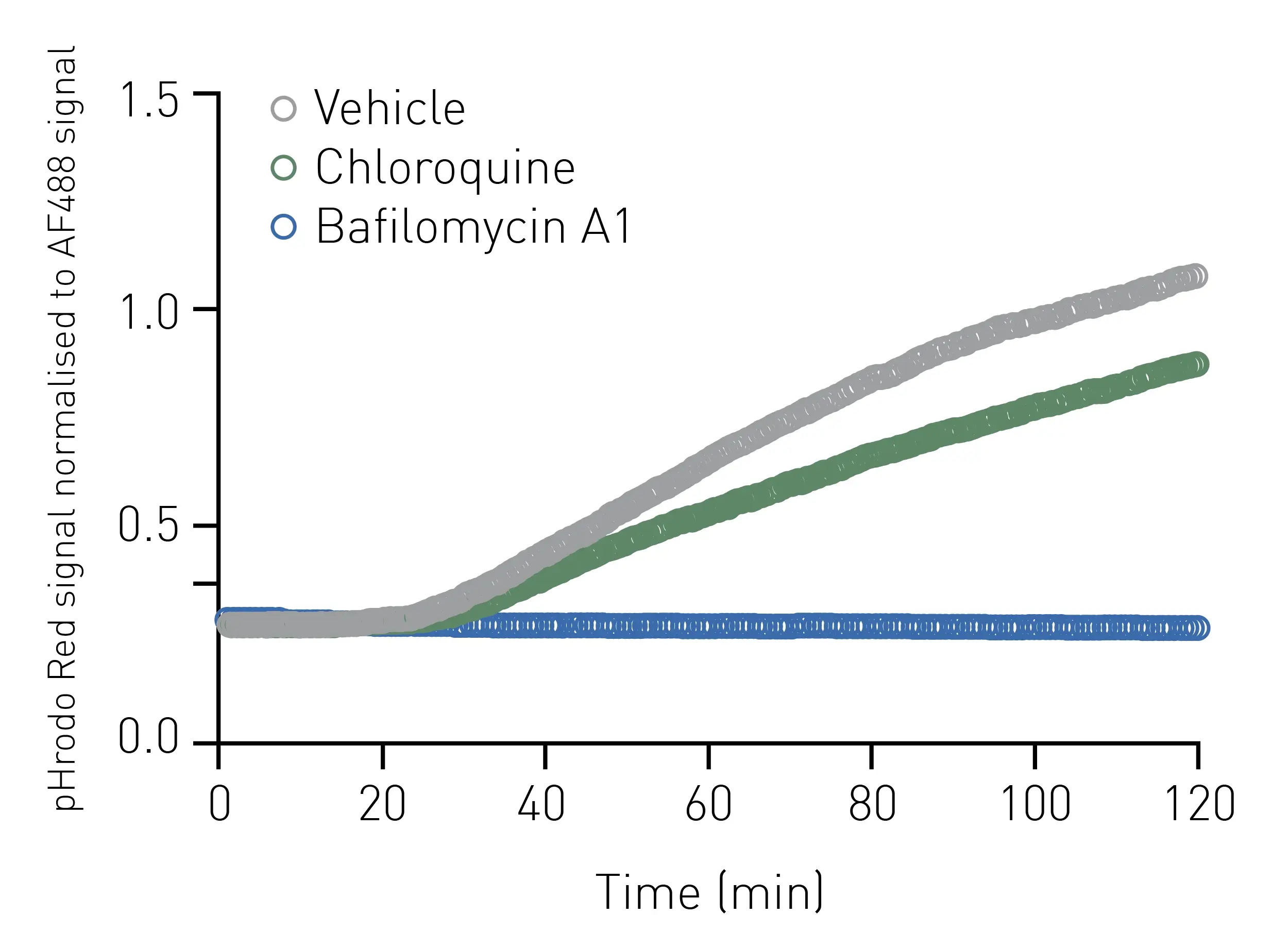
 To account for variation in the uptake of beads between samples, pHrodo Red signal was standardised to AF488 fluorescence which is constitutive and should not change over time (Fig 3).
To assess the biological relevance of pHrodo Red fluorescence as a read out of phagosome acidification, cells were pre-treated with the v-ATPase inhibitor
Bafi lomycin A1 as a negative control5. This completely inhibited phagosomal acidification detected by pHrodo Red coupled beads (Fig 3). Chloroquine, which concentrates in acidic organelles and inhibits the function of key enzymes6, also partially reduced phagosome acidification over time.
Conclusion
pHrodo Red conjugated to carboxylated silica beads, following phagocytosis by macrophages, successfully emitted fluorescence that corresponded to phagosome acidification over time. The pHrodo Red signal was able to be robustly read by BMG LABTECH technologies. Temperature and gas control thereby provide physiological conditions over the entire period of the kinetic measurement. The ability to conduct this assay in a 96-well format allows for significant savings in cell numbers, reagents and cost. Fast speed of acquisition facilitates robust kinetic curves to be plotted and many samples to be processed simultaneously
References
-
Kinchen, J.M. and K.S. Ravichandran, Phagosome maturation: going through the acid test. Nat Rev Mol Cell Biol, 2008. 9(10): p. 781-95.
-
Rock, K.L., H. Kataoka, and J.J. Lai, Uric acid as a danger signal in gout and its comorbidities. Nat Rev Rheumatol, 2013. 9(1): p. 13-23.
-
Yates, R.M. and D.G. Russell, Real-time spectrofluorometric assays for the lumenal environment of the maturing phagosome. Methods Mol Biol, 2008. 445: p. 311-25.
-
Bilkei-Gorzo, O., et al., The E3 ubiquitin ligase RNF115 regulates phagosome maturation and host response to bacterial infection. EMBO J, 2022. 41(23): p. e108970.
-
Wang, R., et al., Molecular basis of V-ATPase inhibition by bafi lomycin A1. Nat Commun, 2021. 12(1): p. 1782.
-
Al-Bari, M.A.A., Targeting endosomal acidification by chloroquine analogs as a promising strategy for the treatment of emerging viral diseases. Pharmacol Res Perspect, 2017. 5(1): p. e00293.