Introduction
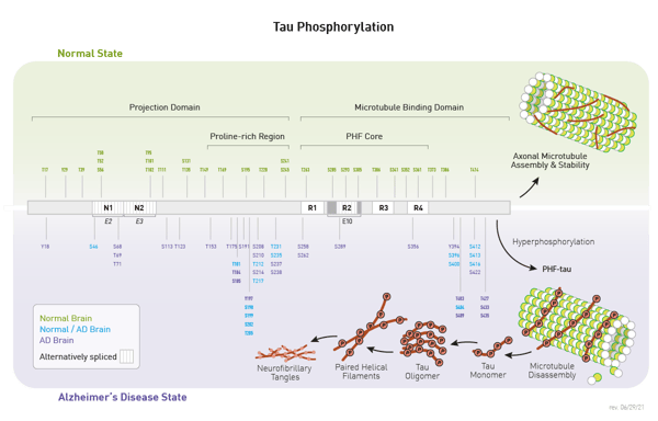
Tau was originally identified for its role as a microtubule associated protein. It has since been shown to be important in axonal growth and function due to its influence on neuronal microtubule stability. The function of tau is regulated by post-translational modification at more than 50 sites. Altered amount of phospho-tau is included in these regulatory modifications (Figure 1).

Tau is of significant clinical and pharmacological research interest because it is among the main components in the neurofibrillary tangles characteristic of Alzheimer’s disease (AD)1. Subsequently it was discovered that high levels of phospho-tau are present in the disease state as well (Figure 1). The precise relationship among tau phosphorylation, tau aggregation, and disease progression is still under investigation.
Here we show the results of detection, by ELISA, of two phospho-tau modifications at threonine 181 and 217. Both have been proposed as functional biomarkers to discriminate between healthy state and AD2. Studies with tau mutations that mimic phospho-tau at Thr217 have shown that it increases phospho-tau at additional sites and increases fibrilization3.
Materials & Methods
- Novus Biologicals: Human Adult Whole Alzheimer’s Brain T issue Lysate (NB820-59363) and Human Adult Whole Normal Brain Tissue Lysate (NB820-59177)
- FastScanTM Phospho-Tau (Thr181) ELISA Kit #58537 and PathScan® Phospho-Tau (Thr217) Chemiluminescent Sandwich ELISA Kit #87749 (Cell Signaling Technology)
- CLARIOstar Plus
Experimental Procedure
- Lysates provided at a 5 mg/mL concentration were diluted in the included FastScan or PathScan buffer
- A 12 point 2-fold dilution series was prepared for each lysate. For FastScan starting lysate concentration is 0.5 mg/ml and 50 µL was used per well. For PathScan starting concentration is 1 mg/mL and 25 µL was used per well.
- ELISA experiments were carried out according to kit instructions found here:
Instrument Settings for PathScan Phospho-Tau (Thr217) Chemiluminescent Sandwich ELISA Kit #87749
|
Optic settings
|
Luminescence
|
|
| Emission |
No filter |
|
|
Gain
|
EDR | |
| Focal height | 11.0 mm | |
|
General settings
|
Optic used | Top |
| Meas. Int. time |
1.00 s
|
|
Instrument Settings for FastScan Phospho-Tau (Thr181) ELISA Kit #58537.
|
Optic settings
|
Absorbance Spectrum | |
| Wavelength range |
400-700 nm |
|
|
Step width
|
2 nm | |
| General settings | Flashes / well |
100
|
|
Settling time
|
0.5 s | |
Results & Discussion
FastScan Phospho-Tau (Thr181) ELISA Kit #58537 results are achieved with a single washing step due to the use of one antibody that is tagged so that the entire antibody and analyte complex is immobilized to the well. In the detection step, collecting spectral data, is useful to trouble-shoot any unexpected results.
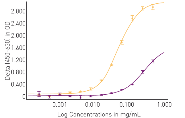
The results shown in figure 2 support that the phospho-tau at Thr181 can be used to discriminate between normal and AD brain tissue samples.
 Absorbance measurements at 630 nm were subtracted from those at 450 nm. The result was plotted against the protein concentration of the tissue samples from normal (purple) and AD (yellow) brains. A 4-parameter fit curve conforms well to the data (R2 = 0.99 for both tissues).
We next tested the PathScan Phospho-Tau (Thr217) Chemiluminescent Sandwich ELISA Kit #87749. This kit can be employed with smaller sample size due to the increase in signal and sensitivity possible with a luminescent assay.
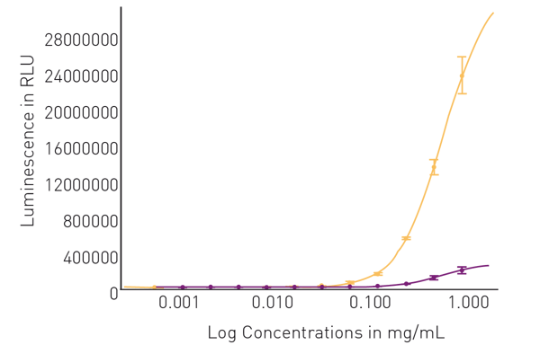
Figure 3 shows high dynamic range of luminescent signal, which is especially evident in high concentration samples of AD tissue.
 Luminescence was plotted against the protein concentration of the tissue samples from normal (purple) and AD (yellow) brains. A 4-parameter fit curve conforms well to the data (R2 = 0.97 or higher for both tissues.
Conclusion
Both the FastScan Phospho-Tau (Thr181) ELISA Kit #58537 and PathScan Phospho-Tau (Thr217) Chemiluminescent Sandwich ELISA Kit #87749 from Cell Signaling Technology generate the expected results in control and experimental human brain tissue samples. In agreement with previously reported literature, both tau phosphorylation sites functionally distinguish normal from AD tissue. Thr217 appears to be somewhat superior to Thr181 as a potential biomarker for AD. CLARIOstar technologies are advantageous for detecting spectral absorbance data and high dynamic range luminescence to ensure optimal performance of these ELISA kits.
References
- Wegmann, S., et al. A current view on Tau phosphorylation in Alzheimer’s disease. Curr. Opin. Neurobiol. (2021) 69: 131- 138
- Thijssen, E.K., et al. Plasma phosphorylated tau 217 and phosphorylated tau 181 as biomarkers in Alzheimer’s disease and frontotemporal lobar degeneration: a retrospective diagnostic performance study. Lancet Neurol. (2021) 20: 739-752
- Wang, X, et al. T217-Phosphorylation Exacerbates Tau Pathologies and Tau-Induced Cognitive Impairment J. Alzheimers Dis. (2021) 81: 1403-1418.

