Introduction
Exosomes are extracellular vesicles (EVs) of endosomal origin. These vesicles contain proteins, lipids, and nucleic acids and facilitate intercellular communication between different cell types within an organism. The rapidly expanding EV research field benefits tremendously from tools for the detection of exosomes and their content. Tetraspanin proteins such as CD9, CD63, and CD81 with exposed domains are particularly enriched in the exosome membrane and are often used as exosome biomarkers. These tetraspanins show a distinct pattern of prevalence on exosomes in different cell types. The Time Resolved Immunoflourescence (TRIFic™) exosome detection kits from Cell Guidance Systems have been developed specifically for analyzing the levels of these tetraspanin proteins on the surface of exosomes.
TRIFic™ exosome detection kits enable researchers to directly measure tetraspanins present on exosomes from sources such as cell culture media, urine, or blood with low background signals and high sensitivity. Using a very sensitive Time-Resolved Fluorescence (TRF) plate reader ensures optimal assay performance. The kit has been evaluated on the FLUOstar Omega, CLARIOstar Plus, and the PHERAstar FSX with all instruments meeting the sensitivity requirements for the kit.
Assay Principle
Step 1: Biotinylated antibody is bound to streptavidin coated assay plates. The kit provides a 96-well microplate with an 8-well strip format.
Step 2: Biological samples are added. Exosomes and any free antigen are captured by the antibody.
Step 3: Europium (Eu)-labelled antibody (identical to antibody used in Step 1) is added and binds specifically to target exosome antigen. The epitopes of any bound monomers are already occupied and not detected. Samples are then read on a Time-Resolved Fluorescence (TRF) plate reader such as the PHERAstar FSX.
Materials & Methods
- TRIFic™ Exosome Detection Kit (Cell Guidance Systems)
- Exo-spin™ Exosome Purification Kit (Cell Guidance Systems)
- Streptavidin coated 96-well microplate (incl. in kit by Cell Guidance Systems)
- TRF microplate reader - PHERAstar FSX (BMG LABTECH)
Exosome Sample Preparation
25 µg of exosomes were purified from conditioned cell culture media using the Exo-spin™ exosome purification kit. The protein amount was determined by Bradford assay. Serial dilutions in PBS by a factor of 0.5 were prepared from 100 µg/ml to 0.8 µg/ml, obtaining a total of 8 different exosome concentrations.
TRIFic™ protocol
100 µl of freshly diluted biotin-conjugated antibody (2 ng/µl) was added to each well of the streptavidin coated plate. Subsequently, incubation and washes using the protocol described below were performed. 100 µl of each exosome dilution (prepared by serial dilution as described above) was transferred to each well. 100 µl of PBS was used as blank. Samples were incubated and washed again as described below. 100 µl of freshly diluted Eu-labeled antibody (0.25 ng/µl) was then added to each well of the streptavidin coated plate. Samples were incubated and washed again as described below. 100 µl of Europium Fluorescence Intensifier solution was then added to each well. The plate was incubated for 15 min at room temperature (RT) on a plate shaker at 750 RPM. TRF was then measured using a TRF compatible microplate reader (PHERAstar FSX).
Incubation and wash protocol
The plate was incubated for 1 hour at RT on a plate shaker at 750 RPM. Each plate was washed three times using 250 µl wash buffer for each wash cycle. The remaining wash buffer was removed.
Instrument settings
|
Optic Settings
|
Time resolved fluorescence (TRF)
|
|
|
Filters:
|
Excitation: 337 nm
Emission: 615 nm
|
|
|
General settings
|
Number of flashes: | 200 with flash lamp |
|
Settling time:
|
0.3 s | |
|
Integration start:
|
200 µs
|
|
| Integration time: | 400 µs | |
Results & Discussion
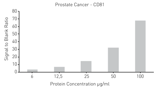
Exo-spin™ isolated exosome samples from conditioned cell culture media were analyzed using the TRIFic™ assay. The expression of CD9, CD63, and CD81 (commonly used surface exosome markers), were measured on exosomes derived from two different cancer cell lines. The data below was obtained using the PHERAstar FSX, however, all three BMG LABTECH instruments exceeded the required performance criteria for the assay.
The error bars for each datapoint in Fig. 2 and Fig. 3 show a very small variance in signal, unnoticeable for some data points. This demonstrates the great consistency and reproducibility offered together by the TRIFic™ kit and PHERAstar FSX. Moreover, the R2 values for every serial dilution show excellent linearity, this is consistent with the data for total protein concentration.

 The data presented on Fig. 4 and Fig. 5 further demonstrates the consistency of the TRF signal generated by the assay over a wide intensity range.
Conclusion
The TRIFic™ assay has been used successfully, together with the BMG LABTECH plate readers. They obtained accurate and reliable TRF signal for different exosome markers. The great linearity and consistency of the signal over a wide range of exosome concentrations demonstrates the robustness of the assay to generate useful data points over a wide dynamic range. This highly sensitive assay can also be used with unpurified samples.
References
- Marina Colombo et al. Annu. Rev. Cell Dev. Biol. (2014) 30: 255-89. DOI: 10.1146/annurev-cellbio-101512-122326
- Diedrik Duijvesz et al. Int. J. Cancer (2015) 137: 2869-78. DOI: 10.1002/ijc.29664
- www.cellgs.com


