Introduction
A large variety of molecules act as ligands for G-protein coupled receptors (GPCRs) physiologically and these receptors are targets for many drugs that are used therapeutically 1. Genome/transcriptome sequencing has enabled the discovery of many novel genes encoding GPCRs in humans and other animals. Identification of the ligands for these ‘orphan’ receptors requires experimental testing of candidate molecules 2,3. To accomplish this, candidate ligands are tested for their effects on cells expressing an orphan GPCR and if a ligand is identified then the orphan GPCR is said to have been ‘deorphanised’. As part of a broader investigation of the evolution of neuropeptide signalling, Ca2+-induced luminescence can be measured in Chinese Hamster Ovary cells (CHO-K1 cells) transfected with orphan GPCRs to test candidate peptide ligands. In recent studies, neuropeptides that act as ligands for many GPCRs in the common European starfish Asterias rubens were identified. For example, the identification of neuropeptides that act as ligands for an expanded family of GPCRs in A. rubens that are related to the human kisspeptin receptor was recently reported 4. One of these receptors (ArKPR6) is activated by the neuropeptide S2 (SGPYSFNSGLTF-NH2), which was one of the first neuropeptides to be identified in starfish 5. Using this as an example of neuropeptide receptor deorphanisation, this application note describes how, by measuring Ca2+-induced luminescence in CHO-K1 cells transfected with ArKPR6, the peptide S2 was identified as a ligand for the orphan GPCR ArKPR6.
Assay principle
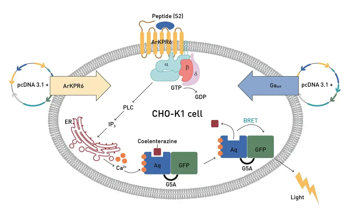
CHO-K1-cells stably expressing a Ca2+-sensitive Green Fluorescent Protein (GFP) - Aequorin fusion protein (G5A) are used as an expression system 6. These cells are co-transfected with an orphan GPCR and a promiscuous G-protein (e.g. Gαqi4, supplied by Luis Alfonso Yañez Guerra, University of Southampton) that couples a large variety of GPCRs to the Phospholipase C (PLC)/ Inositol-1,4,5-tri-phosphate (IP3)/Ca2+ signalling pathway 7. In the presence of the co-factor for aequorin (coelenterazine), binding of a ligand to the orphan GPCR triggers Ca2+-induced luminescence, which is measured. By testing the ligand at different concentrations, a concentration-response curve can be generated.
Materials & methods
- 96-well, white opaque, flat bottom, tissue culture plates (Falcon® 325296)
- Invitrogen Lipofectamine® 3000 Transfection Kit 0.75 ml and Coelenterazine h
- VANTAstar® (BMG LABTECH)
Experimental Procedure
cDNA encoding the orphan GPCR ArKPR6 was custom synthesized with a 5′ partial Kozak translation initiation sequence (CACC) and incorporated into the eukaryotic expression vector pcDNA 3.1 (+) (GenScript, Piscataway, NJ, USA).
CHO-K1 cells stably expressing G5A were cultured in Dulbecco’s Modified Eagle Medium until reaching ~80% confluency. Then cells were transfected with a plasmid containing the orphan GPCR ArKPR6 cDNA and a plasmid containing a cDNA encoding the promiscuous chimeric G-protein Gαqi4.
The peptide S2 (synthesized by Peptide Protein Research Ltd, Fareham, UK; purity > 95%) was dissolved (at 1 mM) in 10% DMSO to improve solubility and then was diluted to a range of concentrations from 10-15 to 10-4 M in basal media. The diluted samples were loaded into white opaque flat bottomed 96-well tissue culture plates, in triplicate (50 µL) for each concentration. Transfected CHO-K1 cells were constantly stirred at 400 rpm at 37°C using the built-in magnetic stirrer heating plate of a VANTAstar multimode-microplate reader (BMG LABTECH) and cells (50 µL) were injected into each well of the plate from the magnetic stirrer, with the reagent injector of the VANTAstar, and then luminescence was recorded for 35 s.
Instrument settings
|
Luminescence, well mode kinetic
|
||
|
Optic settings
|
Filters
|
No filter
|
|
General settings
|
Integration time
|
0.0 s
|
|
Kinetic settings
|
Number of intervals |
35 |
|
Interval time
|
1.00 s |
|
|
Focal height
|
Autofocus |
|
|
Dynamic range
|
EDR |
|
|
Incubation
|
37 °C |
|
Results & Discussion
Figure 2 shows that, by comparison with 10% DMSO and basal media, S2 triggers luminescence in CHO-K1 cells co-transfected with the orphan GPCR ArKPR6 and Gαqi4. Triton X-100 is used as a positive control in these experiments because it non-specifically triggers intracellular Ca2+ elevation in cells and thereby demonstrates cell viability if luminescence is observed. The luminescence measurements with S2, basal media and 10% DMSO were analysed by one-way ANOVA followed by Dunnet’s multiple comparison test, demonstrating a statistically significant increase in luminescence with S2 by comparison with basal media and 10% DMSO.
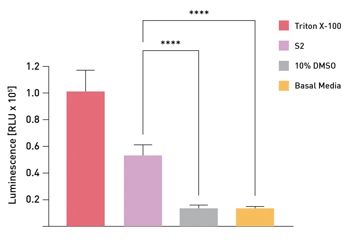
Figure 3 is a concentration-response curve for S2 (10-15 M to 10-4 M), showing that S2 triggers concentration-dependent Ca2+ elevation in CHO-K1 cells co-transfected with the orphan GPCR ArKPR6 and Gαqi4. Furthermore, the EC50 value for this curve (6.24 x 10⁻⁹ M) demonstrates that S2 is a potent ligand for the orphan GPCR ArKPR6. Testing of 10% DMSO diluted to achieve the same concentrations of DMSO as in 10-8 – 10-4 M S2, demonstrates the specificity of the effect of S2. 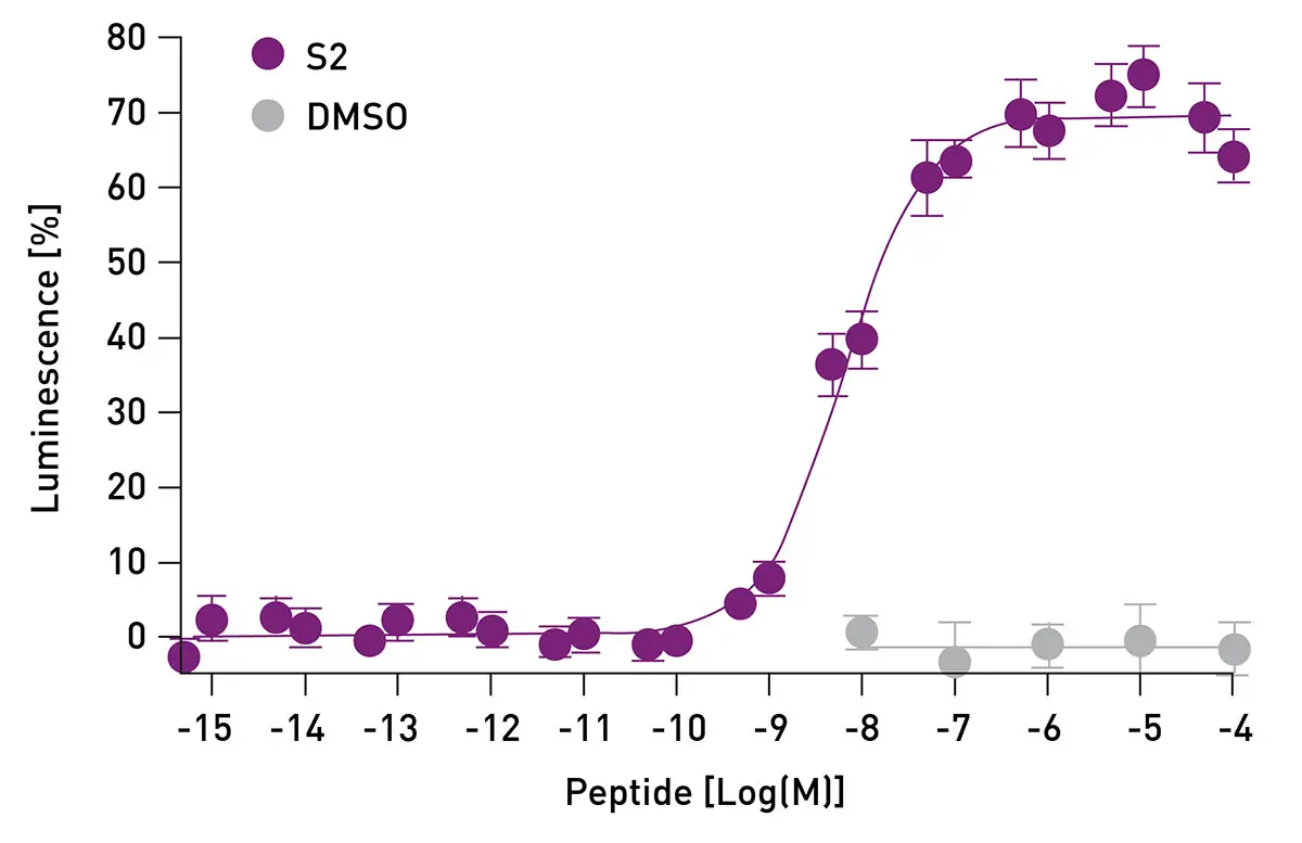
Conclusion
Receptor deorphanisation is providing new insights into the evolution, physiological roles and therapeutic potential of orphan GPCRs 3. Using cell-based assays with promiscuous/chimeric G-proteins, ligands for a huge variety of GPCRs can and have been identified. The VANTAstar multimode-microplate reader combined with a reagent injector compartment that includes a built-in magnetic stirrer heating plate is ideally suited for performing cell-based assays that enable GPCR deorphanisation and enables the necessary manual steps to be reduced and the process to be standardized.
References
- Yang D, et al. Signal Transduction & Targeted Therapy. 2021; 6(1).
- Zhang M, et al. Signal Transduction & Targeted Therapy. 2024; 9(1).
- Hauser AS, et al. Br. J. Pharmacol. 2020; 177, 961-8.
- Escudero Castelán N, et al. BMC Biol. 2022; 20(1), 187.
- Elphick MR, et al. Proc. Roy. Soc. Lond. B. 1991; 243, 121- 7.
- Baubet V, et al. PNAS. 2000; 97, 7260-5.
- Thiel D, et al. eLife. 2024; 12, RP90674.
- Wang Y, et al. Database. 2015; 29, bav038.
- Xiong TC, et al. Front. Plant Sci. 2014; 5, 43.