Introduction
Luciferase-based reporter assays employ insertion of a genetic regulatory element upstream of the luciferase gene. The result is an excellent tool for monitoring gene expression in cells due to wide dynamic range and sensitive detection. The luminescence resulting from expression of the transfected luciferase reporter gene is measured to quantify the activity of the cis-acting or trans-acting components of the biological pathway. In dual luciferase assays, the expression values of one luciferase is usually related to the studied subject whereas the other luciferase reporter is considered as an indicator for transfection efficiency and cell viability.
Assay Principle
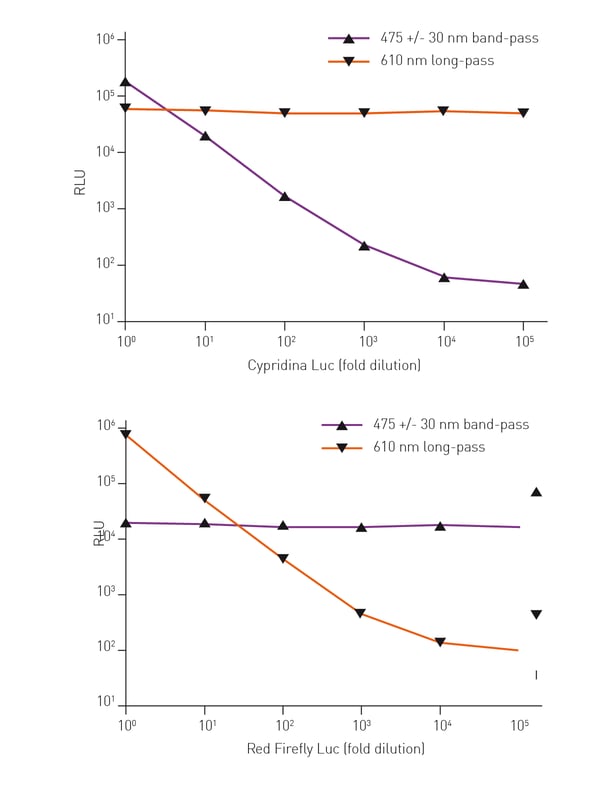
The Thermo Scientific™ Pierce™ Luciferase Dual-Spectral Assays provide a simple one-step detection protocol based on the use of pairs of luciferase enzymes that are spectrally resolved. Therefore their activities can be measured with high sensitivity in the same samples (Figure 1) without the usual quenching step needed to differentiate luciferase signals. Three dual assay systems in which red firefly luciferase is paired with green Renilla, Gaussia, or Cypridina luciferase are available.
 Detecting activity from two spectrally resolved luciferases is a task for which the BMG LABTECH multidetection microplate reader is ideally suited. Light from both reporters can be measured either using the simultaneous dual-emission option which uses two photomultiplier tubes (PMT), or it can be measured sequentially, one emission after the other. Here we demonstrate the sensitivity of the Pierce™ Luciferase Dual-Spectral Reporter Assays, over at least a 100,000-fold luciferase concentration range after normalization to a control reporter, typically red firefly luciferase.
Materials & Methods
- HEK293 stable cell lines expressing Cypridina, Gaussia, Green Renilla, and Red Firefly luciferase under the control of CMV promoter
- Pierce™ Luciferase Cell Lysis Buffer
- Pierce™ Cypridina-Firefly Luciferase Dual-Spectral Assay Kit
- Pierce™ Gaussia-Firefly Luciferase Dual-Spectral Assay Kit
- Pierce™ Renilla-Firefly Luciferase Dual-Spectral Assay Kit
- White, 96-well f-bottom plates (Thermo Scientific)
- BMG LABTECH microplate reader
HEK293 cell lines stably expressing Cypridina, Gaussia, green Renilla, and red firefly luciferases were lysed with Pierce™ Luciferase Cell Lysis Buffer. 1:10 dilutions were prepared in lysis buffer. Analysis of replicates of each serial dilution (10 μL/well) was performed. Cell lysate containing a different second reporter (10 μL/well) was added as a control. For serial dilutions of cell lysate containing Cypridina, Gaussia, or green Renilla luciferase, the second control reporter was red firefly. For serial dilutions of cell lysate containing red firefly, the second control reporter was Cypridina luciferase. The appropriate Pierce™ Luciferase Dual Assay Working Solution was prepared for each assay, and 50 μL/well was injected into each well for a final volume of 70 mL/ well. Luminescence was read through appropriate filters with a 1 second integration time. A more detailed protocol can be found online. An overview of the filter settings is given in table 1.
Table 1: Filter settings for each dual-spectral luciferase assay.
| Assay | Luciferase | Filters | Gain |
| Cypridina-Firefly | Cypridina (Purple graph) | 475 +/- 30 | 2500 |
| Red Firefly | 610 long pass | 2000 | |
| Gaussia-Firefly | Gaussia (Blue graph) | 475 +/- 30 | 2500 |
| Red Firefly | 610 long pass | 2000 | |
| Renilla-Firefly | Renilla (Green graph) | 515 +/- 30 | 2000 |
| Red Firefly | 670 +/- 10 | 3000 |
Results & Discussion
Measuring the separate signals in each of the three pairs of luciferase reporter enzymes in the Pierce™ Luciferase Dual-Spectral Assay Kits was easy, using the appropriate filters (Figures 2, 3 and 4). By selecting filters for the luciferases in each pair that eliminate or greatly reduce interference between them, conditions for each dual-color assay system can be found that provide accurate measurement over 100,000-fold concentration range for each reporter (Figures 2 and 3).
Filter selection is critical for eliminating interference when spectral overlap is present between reporters (Figure 1). Thus for the Renilla-firefly assay, we used a suboptimal red-shifted 670 +/- 10nm bandpass filter for detection of red firefly, which minimizes interference from green Renilla. As a result, interference was only observed when the output from green Renilla was significantly greater than that from red firefly (Figure 4). To separate spectral data in this region, a corrective calculation is needed (see Pierce™ Dual-Spectral Calculator).
Conclusion
These experiments confirm the BMG LABTECH´s microplate reader’s ability to measure the activity of two luciferases that are spectrally resolved. The Thermo Scientific™ Pierce™ Luciferase Dual-Spectral Reporter Assays are sensitive over at least a 100,000-fold dilution range of each luciferase when measured using appropriate filters. Defining the limit of detection in a dual-luciferase assay is difficult and will depend upon luciferase concentration as will the dynamic range.


