Introduction
Staphylococcus aureus infections are associated with high mortality rates. Although often considered an extracellular pathogen, S. aureus can persist and replicate within host cells, evading antibiotics and immune responses, and causing host cell death. Current methods for assessing S. aureus cytotoxicity have focused on identifying exogenous virulence factors that accumulate extracellularly (in the culture media) during bacterial growth, and applied to host cells. Such toxicity tests are limited to the study of extracellular S. aureus and do not capture the phenotypic diversity of intracellular bacteria. To address this issue, Doherty Institute researchers have developed a novel cell toxicity assay named InToxSa, in a 96-well format, to accurately monitor the bacterial toxicity exerted by S. aureus from within host cells1. To achieve this, they took advantage of the ability of S. aureus to invade HeLa cells. Using a combination of gentamicin and lysostaphin antibiotics, extracellular bacteria were eliminated while preserving the viability of intracellular bacteria2. Propidium iodide (PI) was then used as a marker of host cell death.3 The InToxSa assay measures fluorescence changes in HeLa cells over time, reflecting the cell death caused by intracellular S. aureus. The researchers used the InToxSa assay to screen a large collection of S. aureus bacteraemia isolates. The isolates showing a marked reduction in cell death were further characterised using bacterial genomics and evolutionary convergence methods 1. These studies revealed both known and previously undescribed loss-of-function mutations that were significantly associated with decreased intracellular cytotoxicity and increased intracellular persistence.
Assay principle
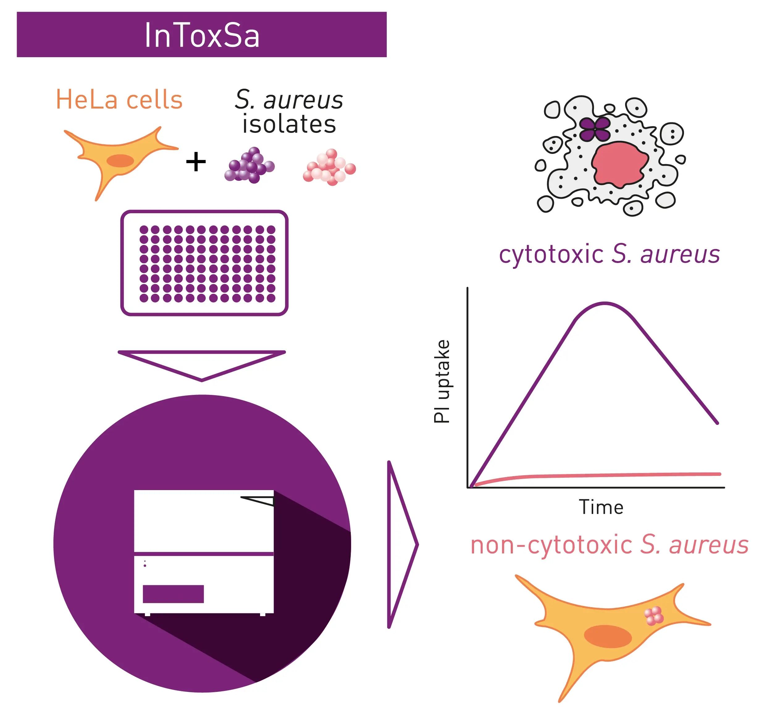
The InToxSa assay continually measures the cell viability of S. aureus-infected HeLa cells over an extended period of time by monitoring propidium iodide (PI) uptake by infected HeLa cells. PI is unable to passively penetrate viable cells with an intact plasma membrane.
Upon HeLa cell death, PI readily diffuses into the cell and strongly binds to its DNA. Once bound to dying HeLa cell DNA, the wavelengths of PI excitation and emission shifts, leading to an increase of fluorescence intensity (Fig. 1).
Materials & methods
- Propidium Iodide (Sigma)
- Gentamicin (Baxter), Lyphostatin (Ambi)
- 96-well plates, clear, PS, Sigma, for bacterial growth
- 96-well plates, black, clear bottom, Sigma, for PI uptake
- BMG Labtech CLARIOstar Plus plate reader with ACU
InToxSa Assay Development
S. aureus isolates were inoculated directly from stabbed frozen plates stock into BHI broth (100 μL), dispensed into flat bottom 96-well plates. Inoculated plates were incubated under agitation (300 rpm) for 16 hours in the CLARIOstar Plus plate reader set at 37°C. Bacterial growth was assessed by OD600 measurement every 10 min. The endpoint optical densities of cultures were used to infer bacterial density (1-unit OD600 corresponding to 5x10⁸ bacteria/ml). Bacterial cultures were standardised and serially diluted in DMEM to reach a multiplicity of infection (MOI) 10. The diluted bacterial suspension (100 µL) was used to infect 40,000 HeLa-CCL2 cells grown in 96-well plates. Infection was synchronised by centrifugation at 500 x g for 10 min at room temperature. Infected plates were incubated for two hours at 37°C and 5% CO2 to allow for S. aureus internalisation.
The infective media was then discarded, and cells washed once with sterile PBS and further incubated for one hour with DMEM (100 μL) containing cell impermeable antibiotics (80 μg/mL gentamicin and 20 μg/mL of lysostaphin) at 37°C and 5% CO2. This treatment was followed by another lower dose (40 μg/ml gentamicin and 10 μg/ml lysostaphin), in media supplemented with 5% fetal bovine serum and 1 μg/mL propidium iodide. Plates were then incubated in the CLARIOstar Plus set at 37°C and 5% CO2 throughout the infection. The fluorescence signal emitted by dying cells was acquired every six minutes from each well for a period of 20 hours. Non-infected control cells were permeabilised with 0.1% Triton X-100 to determine the maximum level of PI uptake by dead HeLa cells and set the fluorescence gain accordingly.
Instrument settings
|
Fluorescence, kinetic
|
||
|
Optic settings
|
Monochromator
|
Ex 535-15
Em 617-20 |
|
General settings
|
Number of flashes
|
50
|
|
Settling time
|
0.5 s |
|
|
Kinetic settings
|
||
|
InToxSa assay
|
No of cycles | 200 |
| Cycle time | 360 s | |
|
Well Scan
|
Spiral averaging mode | |
|
Incubation
|
37°C | |
|
ACU
|
5%, CO2 | |
Results & Discussion
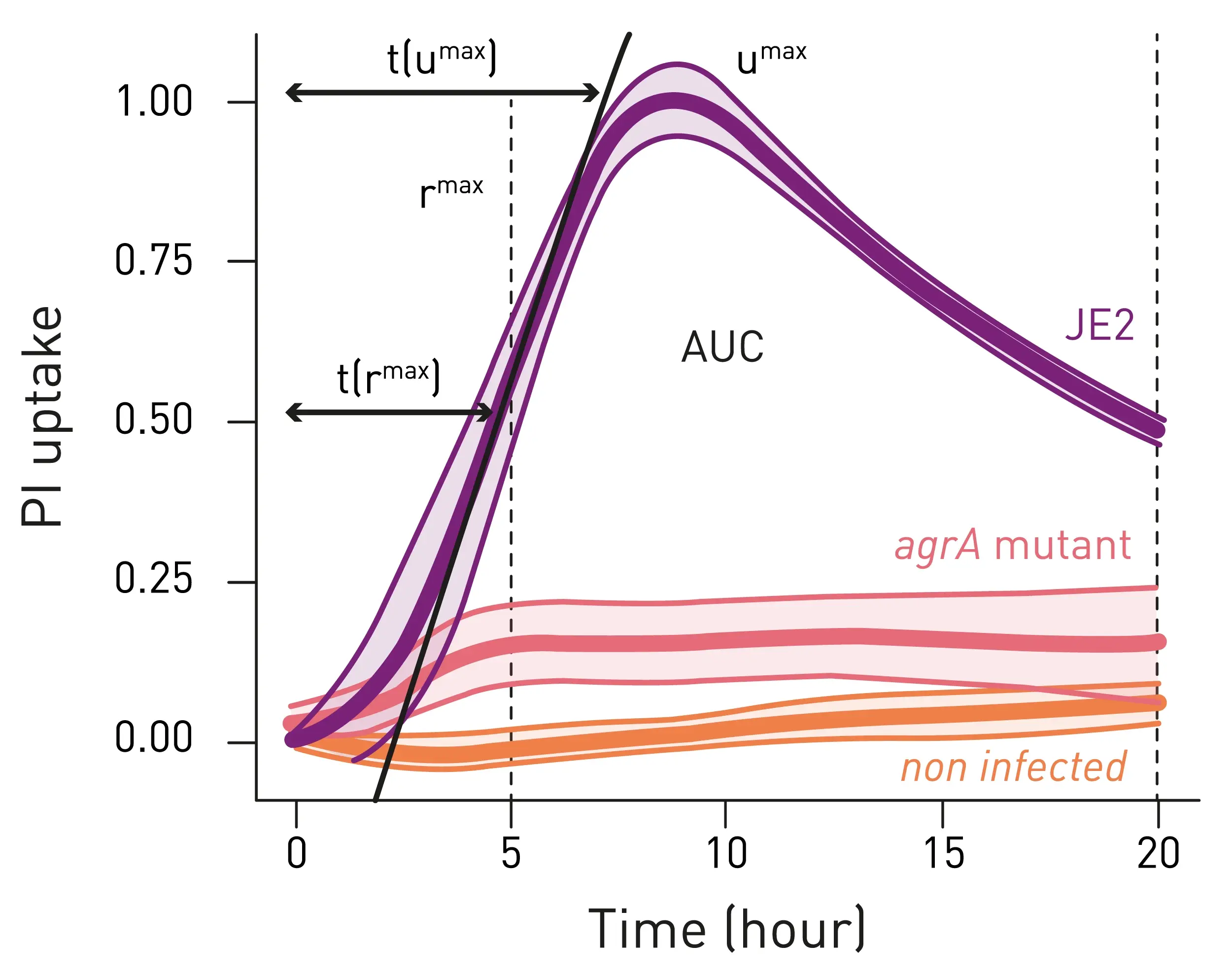
To evaluate the assay’s performance, the intracellular toxicity of wild-type S. aureus strain JE2 was first measured against an isogenic agrA mutant, using non-infected cells as a baseline (Fig. 2). S. aureus JE2 elicited a rapid and substantial increase in PI fluorescence over time, indicative of the well-known high cytotoxicity of this strain (Fig. 2, purple line). In contrast, cells infected with the agrA mutant yielded significantly lower PI uptake (AUC) and a slower PI uptake rate (rmax), suggesting sustained HeLa cell viability during the infection course, in agreement with the reported low cytotoxicity of S. aureus agr mutants (Fig. 2, red line).
The reproducibility of InToxSa outputs was assessed across five experimental replicates, each using both biologically independent HeLa cell culture passages and independent S. aureus cultures. The researchers from the Doherty Institute used regression to fit standardised curves and observed that parameters such as PI uptake, peak PI uptake [μmax] and max. PI uptake rate [rmax] had very low intra-assay variation. These parameters emerged among the most discriminatory and reproducible curve parameters (see paper published in eLife for full statistical data).1
At experimental endpoints, setup was negatively checked for extracellular S. aureus. This indicates that InToxSa exclusively assessed the cytotoxicity caused by intracellular S. aureus. In addition, the InToxSa assay was found to be significantly more sensitive compared to a Trypan blue exclusion assay4, when screening 51 patient S. aureus isolates for reduced cytotoxicity in host cells.
Having established the robustness and sensitivity of this assay, the InToxSa was applied to a panel of 387 clinical isolates of S. aureus bacteraemia. Combined with comparative, statistical and functional genomics, the platform successfully identified mutations in S. aureus clinical isolates that reduced bacterial intracellular cytotoxicity while promoting intracellular persistence. In addition to numerous convergent mutations in the agr quorum sensing system, novel clinical mutations in the gene ausA were also discovered, which encodes the aureusimine non-ribosomal peptide synthetase. These mutations were found to reduce S. aureus cytotoxicity while increasing intracellular persistence. These results show the versatility of the InToxSa assay as a high-throughput cell-based phenomics platform for identifying clinically relevant S. aureus pathoadaptive mutations that promote intracellular residency.
Conclusion
The InToxSa assay represents a reliable and sensitive assay to monitor intracellular bacterial cytotoxicity with high precision. The CLARIOstar Plus is the ideal platform to fulfil the requirements of the InToxSa assay including accurate temperature and atmospheric control to conduct absorbance and fluorescence experiments of long duration.
References
- Hachani A., et. al.; A high-throughput cytotoxicity screening platform reveals agr-independent mutations in bacteraemia associated Staphylococcus aureus that promote intracellular persistence. Elife, 2023 Jun 8:12:e84778.
- Kim JH, et. al.; Alternative enzyme protection assay to overcome the drawbacks of the gentamicin protection assay for measuring entry and intracellular survival of Staphylococci. 2019, Infection and Immunity 87:e00119-19.
- Chitolie M.; Toescu E., High-Throughput method for dynamic measurements of cellular viability: BMG Labtech Application note No. 159.
- Giulieri SG.; Genomic exploration of sequential clinical isolates reveals a distinctive molecular signature of persistent Staphylococcus aureus bacteraemia. 2018, Genome Medicine 10:65.