PHERAstar FSX
Powerful and most sensitive HTS plate reader


Barry Whyte is Application Scientist and Science Writer at BMG LABTECH in the United States. He has PhD and Bachelor of Science (BSc) degrees in biochemistry from the University of Bristol in the United Kingdom and more than 20 years of experience in the life sciences and science communications. Over the years, Barry has worked on three continents and traveled widely. He enjoys building on his international work experience and learning new ways to help scientists advance their research.
The minimum inhibitory concentration or MIC is a crucial metric often used to gauge the effect of antimicrobial agents on microorganisms. In this blog, we take a look at MIC, what distinguishes it from other metrics, trends in the way it is measured, the factors that affect it, and how it can be measured using microplates.
The minimal inhibitory concentration or MIC is a crucial analytical parameter often used in the control of microbial growth by antimicrobial agents. The MIC is typically defined as the lowest or minimum antimicrobial concentration that inhibits visible microbial growth in artificial media after a fixed incubation time and is often expressed in micrograms per milliliter (μg/mL) or milligrams per liter (mg/L). 1,2 It is not the only metric used to look at the effects of antimicrobials on microorganisms. The inhibitory concentration associated with 50% effect (IC50) 2 and the minimal bactericidal concentration , the lowest concentration of an antimicrobial drug that is bactericidal, are also used in different scenarios.3
MIC values provide researchers with a handle on the efficacy of different intervention agents to control microbial growth. As such they have many applications in microbiology, studies of the environment, biotechnology, drug discovery and other areas of research.
While it finds use across microbiology and in other areas of the life sciences, MICs are often cited as metrics in the search for much needed solutions to antimicrobial resistance. Antimicrobial resistance or AMR is a burgeoning global challenge facing society.4 It is ranked as one of the top ten threats to public health worldwide by the World Health Organization and researchers are urgently looking for new solutions to tackle drug-resistant microbes. The measurement of MIC values provides scientists with a way to quantitate the impact of the different agents against microbial growth and the effectiveness of new approaches designed to ameliorate drug resistance in microbial communities. It is also useful for quantifying the effects of antimicrobials on biofilms, communities or arrangements of microorganisms that may also contribute to recalcitrant infections.5
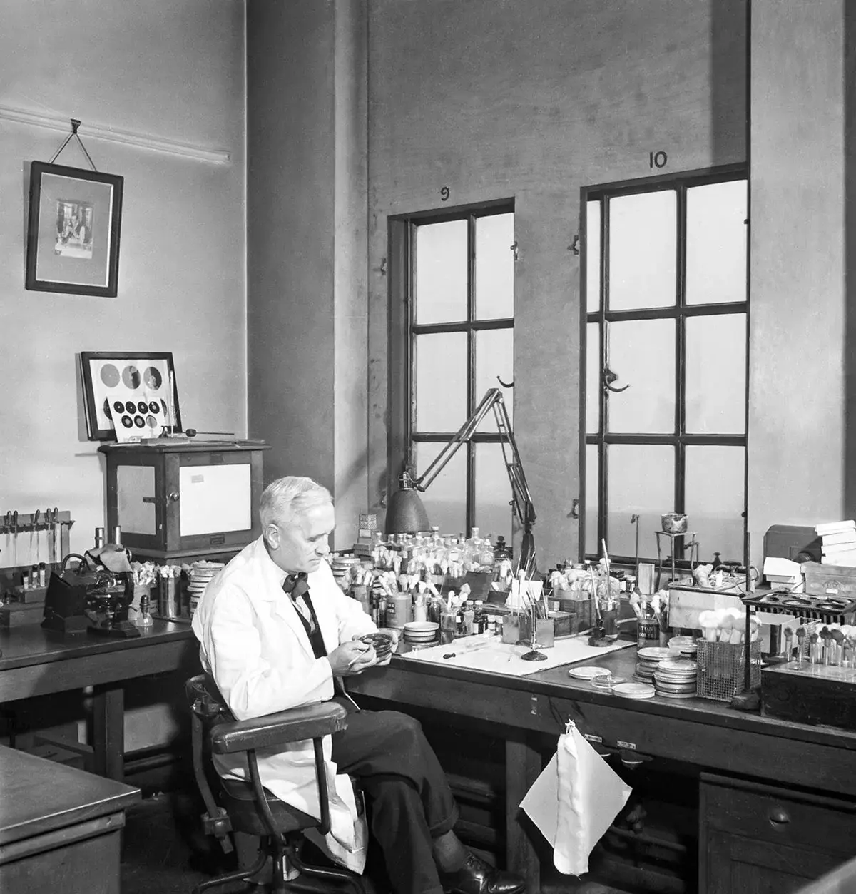
Historically, Alexander Fleming’s early work has been described as a forerunner of contemporary methods to determine MICs (Fig. 1). The discovery of penicillin in 1928 served as a foundation for the quantification of antibacterial activity. His observation that there was a substance that inhibited the growth of Staphylococcus bacteria highlighted that drugs could be found or developed as antimicrobial agents. Fleming’s work was one of the first investigations to quantify antibacterial activity, a concept central to the determination of MICs, and involved the use of serial dilutions to determine the lowest concentration of microbial agent that could inhibit bacterial growth.
Serial dilutions have been widely adopted as a method used to determine MICs. In this approach, a series of decreasing concentrations of antimicrobial agents is tested for its ability to inhibit the growth of a microorganism (Fig. 2). A series of tubes is prepared, each containing a fixed volume of sterile growth medium. The first tube receives the stock potential antimicrobial agent at the highest concentration being tested. A series of dilutions is prepared (for example twofold, tenfold or other appropriate dilution depending on the activity of the agent being tested). This is typically achieved by aliquoting a fixed volume from the first tube to the next tube containing the same volume of fresh medium and repeating the process for the next tubes. The outcome is a geometric progression of concentrations of the potential antimicrobial agent. Each tube is subsequently inoculated with a known number of microorganisms. The incubations are performed under standard conditions, for example at a set temperature and for a known time. After incubation, each tube is visually examined for microbial growth. The lowest concentration of the antimicrobial agent that shows no visible growth is taken as the MIC (Fig. 2).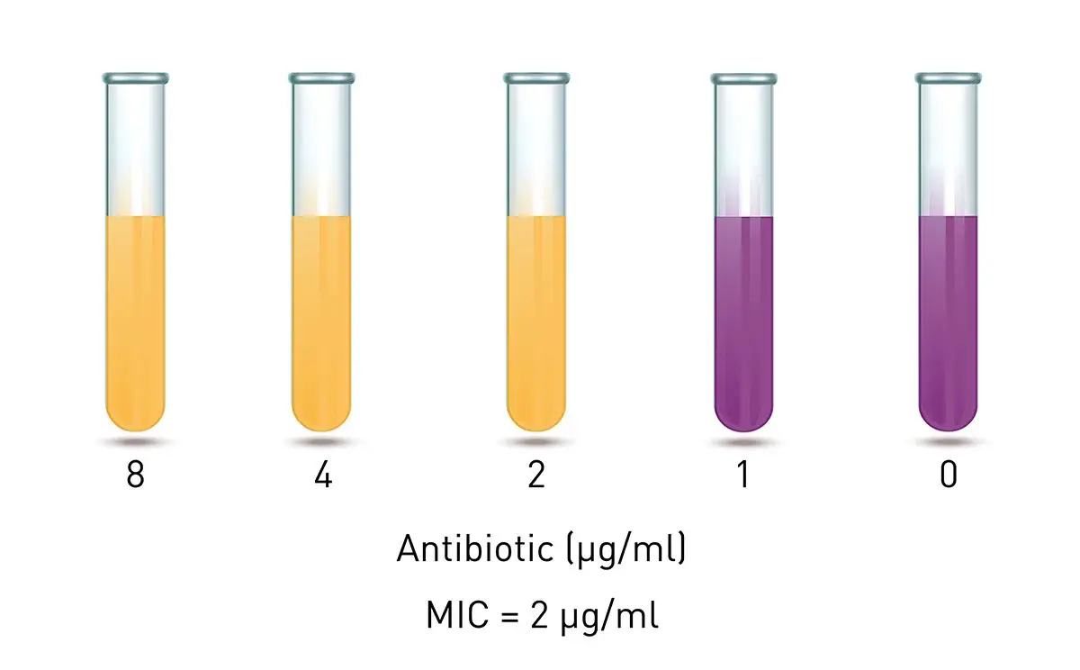 A more recent development for determining MICs is the gradient method. The Etest or Epsilometer test was introduced as a gradient method in the late 1980s for the quantitative measurements of MICs. The Etest is a plastic strip impregnated with a gradient of antibiotic. The strip is placed on an agar plate inoculated with the test organism and the antibiotic diffuses into the agar to create a gradient of concentrations. After incubation under standard conditions, an elliptical zone of inhibition forms around the strip. The point where the edge of the inhibition zone intersects the strip is the MIC value (Fig. 3). In terms of ease of use, gradient methods are much more straightforward than serial dilutions and are fast and applicable to many research settings.
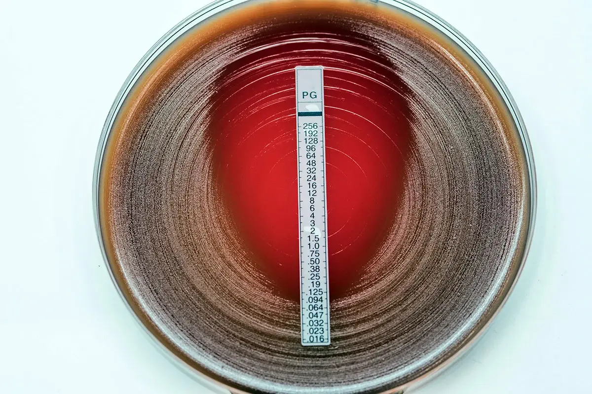
As mentioned earlier, it is essential to perform MIC tests under standard conditions. Careful attention must be given to conditions like pH, temperature and the composition of growth media.
Factors like nutrients, aeration and oxygen may need to be controlled to ensure precise measurements of MICs. Similar considerations may need to be given to moisture and osmolarity.
At the same time, the properties of the antibiotic should also be borne in mind to ensure the fidelity of measurements. For example, molecular structures may inform the way different antibiotics should be solubilized for use in assays (Fig. 4). Small molecules like ampicillin are readily soluble in water or phosphate buffers. Others may require different conditions. Knowledge of the mode of action of different antibiotics can also inform the way experiments are set up for optimal measurements of MIC values.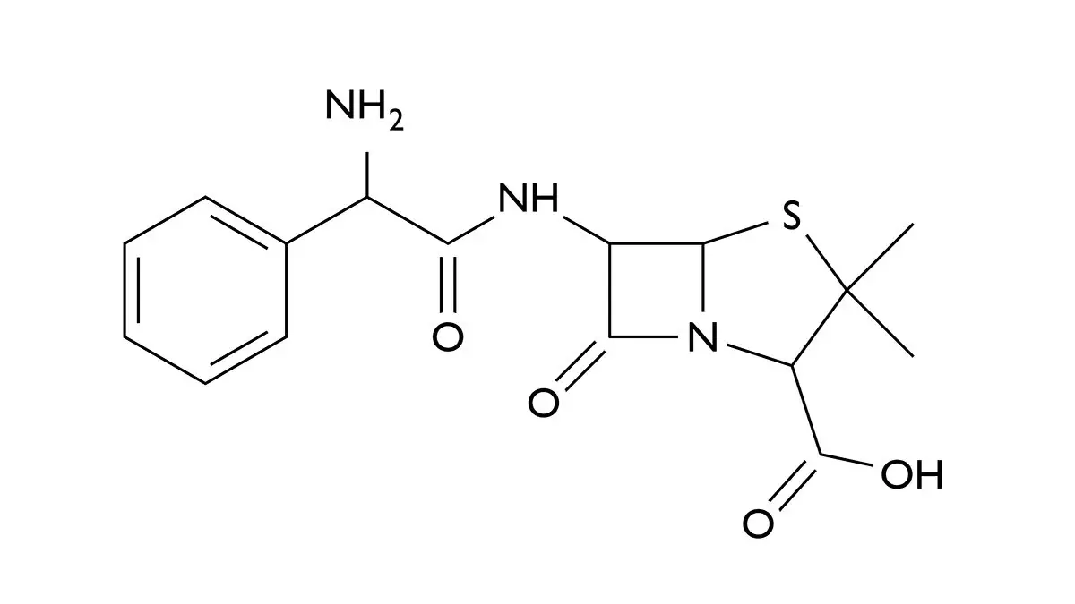 Another important consideration to ensure reliable MIC measurements is to be aware of variability due to different bacterial strains. Different bacterial strains exhibit genetic differences and may employ multiple types of resistance mechanisms. For example, in a study of the “Domestication of Campylobacter jejuni NCTC 11168” it was noted that small differences in the genomes of laboratory reference strains could influence the validity and reproducibility of experimental work.6 The authors quantified differences in 23 reference Campylobacter isolates and compared them with observable differences in common laboratory phenotypes. Bacterial growth assays were performed on an Omega series microplate reader. The impact on the variability of MIC values for ampicillin was one of the endpoints reported. The study highlights the need for careful consideration of genetic variation even within laboratory reference strains.
We saw already how gradient methods accelerated the ability to measure MICs versus other more traditional approaches. Microplate readers in turn offer streamlined solutions that enable a modern approach to monitor MIC of multiple samples (6–1536 wells) in real-time with high accuracy and with no manual intervention. Microplate readers can be used to determine the MIC by measuring the light signal (absorbance, fluorescence or luminescence) of samples in a microplate containing different concentrations of an antibacterial compound. The ability of a microplate reader to measure thousands of samples in a matter of minutes allows for quick and precise determination of the lowest concentration of inhibitor needed. A typical set up for a MIC assay on a 96-well plate is shown in Fig. 5.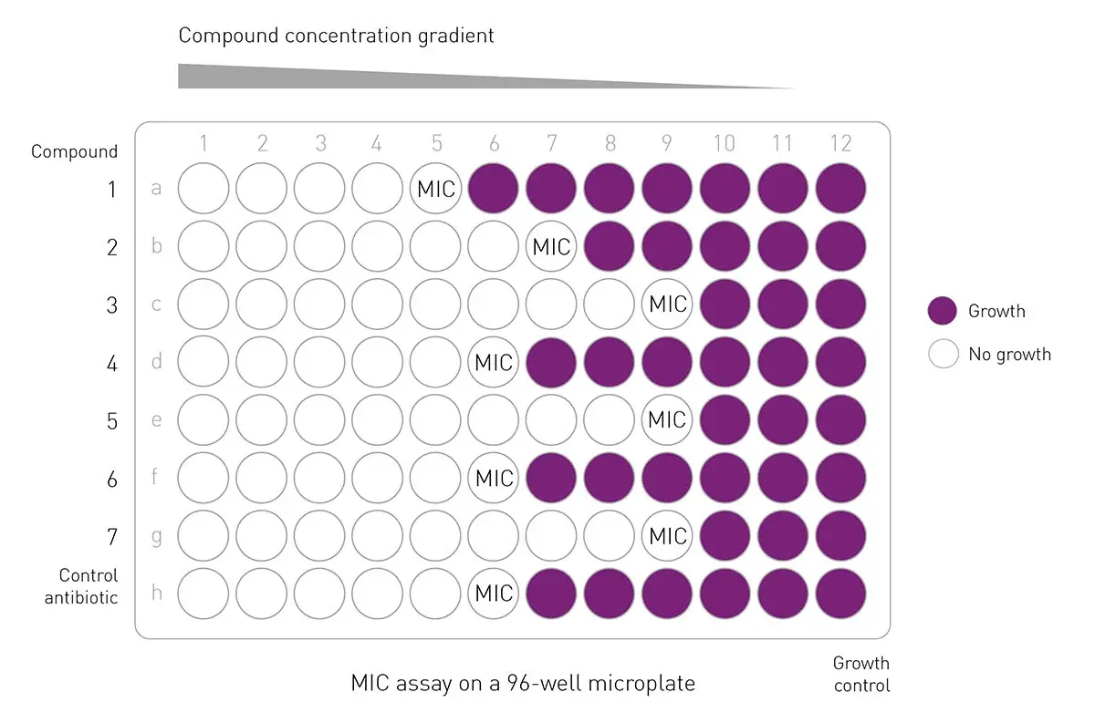
Microplate readers offer several advantages to automate and streamline the measurement of MIC values and to help with interpretation of the results. High throughput measurements are possible on 96-, 384- and 1536-well microplates. This allows for testing of many different antimicrobial concentrations as well as multiple microbial strains in parallel.
Microplate readers automate the measurements of OD600 which is indicative of bacterial growth. You can read more about measuring microbial growth using OD600 in this blog from BMG LABTECH and can find some and can also find possible troubleshooting approaches in the HowTo Note ‘How to optimise OD600 measurements`. The use of microplate readers reduces the potential impact of human error and its associated variability of measurements and leads to more reliable results. Measurements can be made at regular intervals (e.g., minutes or hours) without manual interventions. Real time data can be readily collected which accelerates analysis. In addition to high precision and sensitivity of measurements, microplate readers come equipped with data collection and software analysis options. MIC values can therefore be determined based on defined criteria and graphs plotted to assist in calculations (Fig. 6). 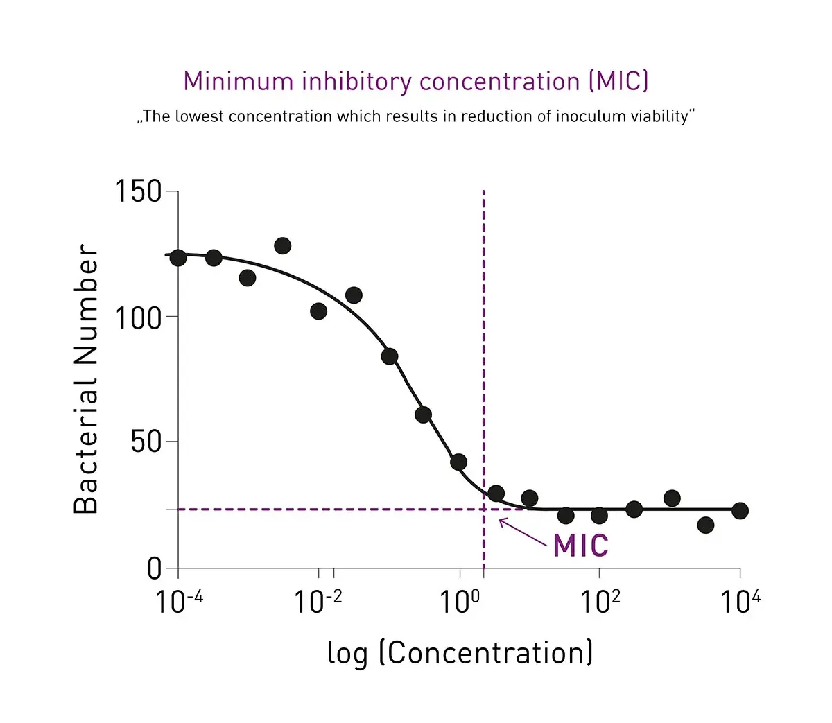 Overall, microplate readers help to standardize the process for determining MIC values and ensure that conditions are consistent across all wells in the microplate. Additional features like incubation, shaking and an Atmospheric Control Unit provide distinct benefits for the support of consistent growth conditions for microorganisms that have more demanding parameters for growth. This type of consistency is essential for accurate determination of MIC values for more fastidious microbes. The Atmospheric Control Unit from BMG LABTECH provides researchers with a system that enables control of both the oxygen and carbon dioxide concentrations in an independent manner. Consistent stirring options also deliver benefits for MIC experiments.
In the application note High-throughput determination of bacterial growth kinetics using a BMG LABTECH microplate reader, the ability to measure growth rates and compare MIC values for biocide-resistant mutants of salmonella and their parent strains was instrumental in testing the actions of different biocides. Bacterial growth curves were monitored in absorbance mode automatically over 24 hours (Fig.7). The exposure to biocides was selected for strains with increased tolerance to microbial agents and it was demonstrated that there was no obvious cost to the fitness of the microorganisms under study. MIC values were crucial parameters measured for the different biocides tested in the study.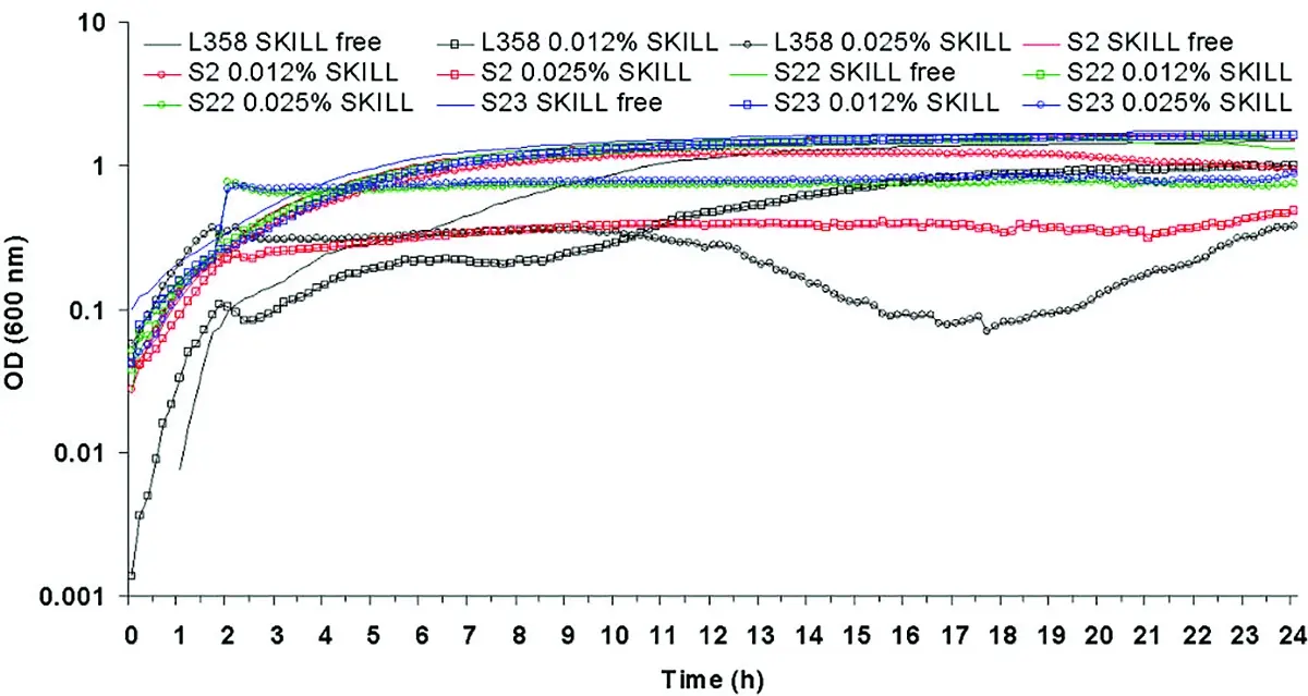 In the application note High-throughput method for dual assessment of antifungal activity and growth kinetics using a FLUOstar Omega MIC values for antimicrobial agents were determined from kinetic measurements. The effects of the antimicrobial agent on yeast growth were measured on a microplate reader that allowed growth and inhibitory effects to be measured in parallel. In the experiments reported MIC assays provided a sensitive and accurate analysis of growth kinetics that demonstrated subtle dose-dependent drug effects that otherwise might be missed by endpoint readings. The benefits included the ability to quickly test a range of different antimicrobial agents at different concentrations on Candida albicans and other pathogens. If different fluorescent strains are available, both growth and inhibitory properties of mixed microbial populations can be assessed in tandem.
The application note “Determination of Minimum Inhibitory Concentration (MIC) of antibiotics using OD600”, highlights how the VANTAstar was successfully used to detect the MIC of two different antibiotics on the growth of E. coli using OD600. The setup in a 96-well plate thereby allowed the evaluation of 10 concentrations in quadruplicate for each antibiotic, increasing throughput significantly.
In the paper “Influence of sub-inhibitory concentrations of antimicrobials on micrococcal nuclease and biofilm formation in Staphylococcus aureus“ a group of researchers used MIC values to look at the impact of antimicrobials on enzyme production related to the biomass of biofilms.7 The work involved the determination of MICs for biofilms using crystal violet assays performed on a BMG LABTECH Omega series microplate reader. Overall micrococcal nuclease activity was shown not to directly influence total biomass of the biofilm but the deletion of the nuc1 gene did stimulate polysaccharide production in the in vitro biofilm assay used in the investigation. Biofilm formation and micronuclease activity in planktonic and biofilm cultures were studied while applying various sub-MICs of antimicrobial agents to three strains of Staphylococcus aureus.
The need for the rapid and accurate determination of MIC values is increasing due to several factors. First MIC values are crucial in evaluating the efficacy of new antimicrobials and other drugs during research and development. Second, the global increase in antimicrobial resistance necessitates precise and effective use of both new and existing antibiotics. MIC values help in selecting the most effective antibiotic options in specific scenarios. They also help in deciding the optimal doses of antimicrobial agent to use versus resistant strains. Collectively, this can assist in slowing the spread of antimicrobial resistance.
While BMG LABTECH microplate readers are not for use in environments that require in vitro diagnostic approved devices and processes, they may provide researchers with many solutions to support fundamental research in non-clinical settings where scientists are looking to use MIC values as part of their research efforts.
Many areas of investigation require robust research into antimicrobials and their modes of action. Overall, the growing threat of antimicrobial resistance and the need for new and more effective treatments underscore the increasing importance of MIC values in research settings across the globe.
What is the preferred BMG LABTECH microplate reader for specific needs and applications related to the determination of MIC values? Absorbance detection for the measurement of OD600, a crucial measurement in the determination of MICs, is available on BMG LABTECH’s complete portfolio of microplate readers with the ultra-fast spectrometer. The exception is the NEPHELOstar Plus which is a dedicated laser-based nephelometer for light scattering and turbidity measurements.
BMG LABTECH also offers a range of multi-mode detection devices for sensitive fluorescence and luminescence measurements.
Bacteria require specific temperatures and aeration for maximum growth rates. To ensure optimal growth conditions, all BMG LABTECH readers offer accurate temperature regulation up to 45°C (optionally up to 65°C). Three shaking modes with adjustable speed up to 700 rpm (optionally to 1100 rpm) provide optimum aeration settings for your strain. Additionally, the VANTAstar, CLARIOstar Plus, the Omega series and the SPECTROstar Nano can be equipped with an extraordinary robust transport system for shaking 24/7 where required.
The VANTAstar, the CLARIOstar Plus, the Omega series and NEPHELOstar Plus can be combined with the Atmospheric Control Unit making them the preferred choice for different kinds of live cell assays including bacterial growth assays.
Both the VANTAstar and CLARIOstar Plus further allow for wavelength flexibility and include Enhanced Dynamic Range technology for superior performance in a single luminescence or fluorescence run. They also offer increased light transmission and sensitivity courtesy of Linear Variable Filter MonochromatorsTM and different filter options.
All BMG LABTECH microplate readers have exceptionally fast reading capabilities. In addition, the Omega series, CLARIOstar Plus, and PHERAstar® FSX microplate readers come with on-board injectors that can offer the very best options for detection at the time of injection. The VANTAstar can be equipped with a modular injection unit. The SPECTROstar Nano comes with a dedicated cuvette-port which can also be used to study bacterial growth over time in a cuvette-based approach.
In his testimonial, Studying bacterial pathogens on BMG LABTECH readers, Andrew Roe from the University of Glasgow discusses the use of BMG LABTECH readers. His work is mainly focused on bacterial pathogens, the different sites of the body where they cause infections, and the genes that are involved in this process.

Collectively, BMG LABTECH multi-mode readers combine high-quality measurements with miniaturised assays, short measurement times, and offer considerable savings on materials and other resources.
The NEPHELOstar Plus offers turbidimetric measurements for the determination of bacterial growth at very high sensitivity. It can be used for example to study the early stages of bacterial growth.
BMG LABTECH microplate readers are available for research purposes only.
Configure your microplate reader and get an initial recommendation!
Powerful and most sensitive HTS plate reader
Most flexible Plate Reader for Assay Development
Flexible microplate reader with simplified workflows
Upgradeable single and multi-mode microplate reader series
Absorbance plate reader with cuvette port
Learn about the bacterial endotoxin test (BET test) and its role in ensuring the safety of pharmaceuticals, biologics and medical devices.
The monocyte activation test is a cell-based assay used to detect pyrogens. Learn more about the monocyte activation test and how microplate readers can support these crucial tests for quality control and bioanalysis.
The recombinant factor C (rFC) test is used to detect bacterial endotoxins that can cause fever and adverse events when introduced into the body. Learn about the rFC test and the advantages it offers for endotoxin detection in the life sciences.
Pyrogen tests are vital to ensure the safety of different health interventions including pharmaceuticals, medical devices and an array of biological products. This blog looks at the different types of pyrogens as well as some of the widely used pyrogen tests.
Learn about applications for bacterial metabolism on a microplate reader.
Gene reporter assays are sensitive and specific tools to study the regulation of gene expression. Learn about the different options available, their uses, and the benefits of running these types of assays on microplate readers.